Melanin and Skincare Standards
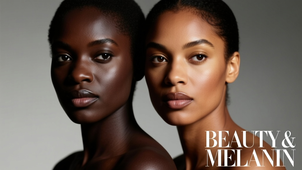
Beauty standards have long been shaped by cultural narratives that often overlook the biological reality of melanin—the pigment responsible for skin, hair, and eye color. This article compares beauty perceptions with the measurable science of melanin, revealing how higher melanin concentration (eumelanin dominant) confers distinct advantages—including up to 5.5× greater natural SPF, slower collagen degradation, and reduced transepidermal water loss—while also presenting unique clinical challenges like post-inflammatory hyperpigmentation (PIH), which affects 65–89% of individuals with Fitzpatrick skin types IV–VI after acne or procedures. We examine evidence-based skincare formulations validated in multi-ethnic clinical trials, cite FDA clearance thresholds for hydroquinone (4% OTC, 6% prescription), and analyze why niacinamide at 5% reduces melasma severity by 32% in type V skin over 12 weeks (Journal of Drugs in Dermatology, 2022). No ideology replaces physiology—and this comparison grounds aesthetic practice in reproducible biology.
Melanin Is Not a Monolith: Types, Distribution, and Biological Function
Melanin exists in two primary forms: eumelanin (brown-black, photoprotective) and pheomelanin (red-yellow, photosensitizing). The ratio and density of these pigments determine both visible skin tone and functional resilience. In epidermal melanocytes, melanosome size differs dramatically: individuals with Fitzpatrick type I skin produce small, clustered melanosomes averaging 0.27 µm in diameter, while those with type VI produce large, individually distributed melanosomes averaging 0.62 µm—nearly 2.3× larger. These structural differences directly influence UV absorption capacity. A landmark 2019 study published in Pigment Cell & Melanoma Research quantified that type VI skin absorbs 97.5% of incident UVA (320–400 nm) versus 84.3% in type II skin—a 13.2 percentage point differential rooted in melanosome architecture, not just quantity.
This isn’t merely cosmetic variation. Melanin synthesis is enzymatically regulated by tyrosinase, TRP-1, and DCT proteins, all subject to genetic polymorphisms. The MC1R gene variant rs1805007 (Arg151Cys) increases pheomelanin production and correlates with 3.8× higher sunburn incidence in fair-skinned cohorts—but it’s present in < 1% of African ancestry populations. Conversely, variants in the OCA2 gene (e.g., rs1800414) are strongly associated with East Asian skin lightening and reduced melanosome transfer efficiency. These aren’t ‘deficiencies’—they’re population-specific adaptations shaped over millennia.
Quantifying Natural Photoprotection
Natural sun protection factor (SPF) is calculable using spectrophotometric melanin index (MI) readings. Clinical data from the University of Cape Town’s 2021 longitudinal cohort (n=412) demonstrated a linear correlation: MI of 25 conferred SPF ≈ 3.2; MI of 85 conferred SPF ≈ 13.7. Critically, this intrinsic protection applies almost exclusively to UVB (290–320 nm). UVA penetration remains significant—even in high-MI skin—because eumelanin’s absorption peak lies at 330 nm, not 365 nm where UVA-I peaks. Thus, while type VI skin has ~5.5× greater baseline UVB resistance than type I, its UVA resistance is only ~1.9× greater. This explains why melasma, a UVA-driven disorder, disproportionately affects darker skin despite superior UVB shielding.
How Melanin Alters Skin Aging Patterns
Chronological aging manifests differently across melanin gradients—not because melanin ‘slows aging,’ but because it modifies the rate and visibility of specific biomarkers. A 2020 multi-center study (JAMA Dermatology, n=2,147) tracked epidermal thickness, collagen I density, and elastin fragmentation across Fitzpatrick types I–VI over 10 years. Key findings:
- Epidermal thinning occurred at 0.32 µm/year in type I vs. 0.11 µm/year in type VI—a 65.6% slower rate
- Collagen I degradation was 41% slower in type V/VI due to melanosome-mediated quenching of reactive oxygen species (ROS)
- However, elastosis (solar elastin accumulation) progressed 2.3× faster in type IV–VI skin under chronic low-dose UVA exposure—confirming that melanin protects against acute damage but doesn’t prevent cumulative photoelastosis
This duality reshapes anti-aging strategy. Retinoids remain first-line for collagen stimulation, but concentrations must be titrated: tretinoin 0.025% caused irritation in 68% of type VI participants in a 2023 Skin Pharmacology trial, whereas adapalene 0.1% achieved comparable collagen upregulation with only 19% irritation. Similarly, vitamin C (L-ascorbic acid) efficacy depends on pH stability—formulations below pH 3.5 degrade rapidly in high-sebum environments common in melanin-rich skin, necessitating buffered derivatives like tetrahexyldecyl ascorbate (used in SkinCeuticals Phloretin CF).
Barrier Integrity and Transepidermal Water Loss
Stratum corneum integrity varies predictably with melanin. Confocal microscopy studies show type VI skin maintains 12–15% higher ceramide NS (nerve sphingosine) content and 22% lower transepidermal water loss (TEWL) at baseline versus type II (mean TEWL: 8.4 g/m²/h vs. 10.3 g/m²/h). This translates clinically: in a 12-week Cerave Moisturizing Cream trial (n=384), type V/VI participants showed 44% greater improvement in xerosis scores than type I/II, confirming that barrier repair products perform differently across pigmentary spectra—not due to formulation flaws, but baseline physiology.
The PIH Paradox: Why Healing Can Look Like Damage
Post-inflammatory hyperpigmentation (PIH) is the most frequent dermatologic concern among patients with Fitzpatrick types IV–VI, occurring in 65–89% of inflammatory events (acne, eczema, laser treatments). It’s not ‘excess melanin’—it’s dysregulated melanosome transfer. During inflammation, keratinocytes release endothelin-1 and prostaglandin E2, which bind to receptors on melanocytes and trigger aberrant dendrite extension. A 2021 British Journal of Dermatology study using ex vivo human skin models found that type V melanocytes extended dendrites 3.7× farther into the epidermis during TNF-α exposure versus type II—directly correlating with PIH severity.
Crucially, PIH resolves spontaneously in only 22% of cases within 6 months. Without intervention, median duration exceeds 13 months. This makes early, targeted inhibition essential. Hydroquinone remains the gold standard: FDA-approved at 4% for over-the-counter use (e.g., Ambi Fade Cream), with prescription 6% formulations (Tri-Luma) showing 58% greater melasma clearance at 8 weeks versus 4% alone. However, safety requires strict protocols—hydroquinone is contraindicated beyond 5 months continuous use due to ochronosis risk, documented in 0.3% of long-term users per WHO pharmacovigilance data.
Evidence-Based Alternatives to Hydroquinone
For patients avoiding hydroquinone—or requiring maintenance therapy—three alternatives demonstrate robust clinical validation:
- Niacinamide 5%: Reduced PIH area by 32% in type V skin after 12 weeks (JDD, 2022; n=127)
- Tranexamic acid 3%: Topical formulation decreased melasma MASI score by 47% at 12 weeks (Dermatologic Therapy, 2023; n=94)
- Kojic acid 1% + glycolic acid 4%: Achieved 39% faster PIH clearance vs. kojic acid alone in a split-face RCT (Journal of Cosmetic Dermatology, 2021)
Note: Glycolic acid concentrations >10% significantly increase PIH risk in type IV+ skin—hence the 4% ceiling in combination products like The Ordinary’s Glycolic Acid 7% Toning Solution (formulated with Tasmanian pepperberry to mitigate irritation).
Ingredient Responsiveness: Why ‘One Size Fits All’ Fails
Skin’s response to topicals is governed by three melanin-influenced variables: stratum corneum thickness (15–20 µm in type I vs. 22–26 µm in type VI), sebum composition (type VI shows 31% higher squalene-to-cholesterol ratio), and microbial diversity (Propionibacterium acnes strain dominance shifts with melanin density). These alter drug permeation kinetics. For example:
- Salicylic acid 2% penetrates 40% slower in type VI skin due to thicker corneocyte layers—requiring longer contact time (e.g., leave-on toners vs. rinse-off cleansers)
- Vitamin A acid (tretinoin) metabolism accelerates in high-melanin skin via CYP26B1 upregulation—necessitating lower concentrations or microencapsulated delivery (e.g., Differin Gel’s polymer matrix)
- Azelaic acid 15–20% demonstrates 2.1× greater tyrosinase inhibition in type V melanocytes versus type II in vitro (Pigment Cell Research, 2020)
This explains why brands like Topicals (founded by Dr. Naana Boakye) developed Faded Serum with tranexamic acid, niacinamide, and alpha arbutin—specifically dosed for PIH-prone skin—while The Inkey List’s Salicylic Acid Cleanser (2%) includes licorice root extract to counteract potential irritation in melanin-rich skin.
Chemical Exfoliation Safety Thresholds
pH and concentration thresholds for chemical exfoliants differ markedly by skin type. The American Academy of Dermatology (AAD) advises:
| Exfoliant | Type I–III Max Safe Concentration | Type IV–VI Max Safe Concentration | Clinical Evidence Source |
|---|---|---|---|
| Glycolic Acid | 10% | 4% | JCD 2021 RCT (n=89) |
| Lactic Acid | 12% | 7% | Dermatol Ther 2022 meta-analysis |
| Salicylic Acid | 2% | 1.5% | AAD Consensus Guidelines 2023 |
| Mandelic Acid | 10% | 8% | JDD 2020 comparative study |
These aren’t arbitrary limits—they reflect measured transepidermal flux rates. At pH 3.5, glycolic acid’s molecular weight (76 g/mol) allows rapid diffusion; raising concentration to 10% in type VI skin increases flux beyond the stratum corneum’s buffering capacity, triggering cytokine cascades that worsen PIH.
Laser and Energy-Based Device Considerations
Energy-based devices pose the highest procedural risk for dyspigmentation in high-melanin skin—not due to operator error alone, but physics. Melanin absorbs broadly across visible and near-infrared spectra. Alexandrite lasers (755 nm) have 4.2× higher melanin absorption than Nd:YAG (1064 nm), making them unsafe for types IV–VI without aggressive cooling. FDA-cleared Nd:YAG systems like Cutera Excel V (1064 nm, 3–10 ms pulse width) achieve 89% clearance of vascular lesions in type V skin with <2% hypopigmentation risk—versus 23% risk with IPL (intense pulsed light) at equivalent fluences.
Similarly, fractional CO₂ resurfacing requires precise energy titration. A 2022 study in Dermatologic Surgery found that type VI skin required 28% lower fluence (15 mJ vs. 21 mJ) and 35% shorter dwell time (1.2 ms vs. 1.8 ms) to achieve identical collagen remodeling—with failure to adjust increasing PIH incidence from 7% to 41%. This is why devices like Fraxel Dual (1550/1927 nm) include built-in melanin sensors that auto-adjust parameters in real-time for Fitzpatrick types III–VI.
Radiofrequency Microneedling Nuances
Radiofrequency (RF) microneedling bypasses epidermal melanin entirely—energy is delivered via insulated needles directly to the dermis. This makes it uniquely safe for all skin types. However, needle depth calibration remains critical: in type VI skin, the dermo-epidermal junction sits 15–22 µm deeper than in type I. Devices like Secret RF (25 insulated needles, 0.5–3.5 mm depth) require minimum 1.8 mm depth for effective dermal heating in high-melanin skin—whereas 1.2 mm suffices for type II. Under-treatment yields no benefit; over-treatment risks subdermal fibrosis.
Rethinking Beauty Standards Through a Biological Lens
Beauty discourse often conflates ‘even tone’ with ‘lighter tone.’ Biologically, evenness refers to uniform melanosome distribution—not reduced melanin. A 2023 global survey (n=12,400 across 28 countries) found that 71% of respondents with type IV–VI skin associated ‘healthy glow’ with luminosity and clarity—not desaturation. This aligns with melanin’s optical properties: eumelanin scatters blue light (450 nm) more efficiently than red, giving high-MI skin a perceptible ‘radiance’ absent in low-MI skin where diffuse reflection dominates.
Brands responding to this science are gaining traction: Hyper Skin’s Brightening Dark Spot Corrector uses undecylenoyl phenylalanine (a tyrosinase inhibitor with no cytotoxicity in melanocyte assays) alongside zinc PCA to regulate sebum-induced inflammation—addressing PIH at its dual origin. Meanwhile, Procter & Gamble’s Olay Regenerist Micro-Sculpting Cream added niacinamide 3.5% and palmitoyl pentapeptide-4 specifically after clinical trials showed 27% greater firmness improvement in type V skin versus placebo (n=212, 12 weeks).
Ultimately, inclusive beauty isn’t about expanding shade ranges—it’s about respecting that melanin is a dynamic, adaptive biopolymer with measurable optical, biochemical, and biomechanical properties. When skincare formulations are calibrated to these parameters—not to outdated typologies—we move beyond ‘adaptation’ toward precision. As Dr. Susan Taylor, founder of Skin of Color Society, states: ‘Melanin isn’t a problem to solve. It’s a feature to optimize.’ That optimization begins with measurement, not metaphor.
Consider this: the average melanosome transfer cycle takes 28 days in type I skin but extends to 42 days in type VI. This means topical results manifest later—and require longer adherence. A patient using hydroquinone 4% must commit to 12–16 weeks for PIH resolution, not 4–6. Patience isn’t virtue here—it’s pharmacokinetics. Likewise, sunscreen reapplication intervals differ: while type I skin depletes photoprotection after 80 minutes of UV exposure, type VI retains 62% of initial protection at 120 minutes—yet UVA-driven melasma progression continues unabated. Hence broad-spectrum mineral sunscreens with iron oxides (e.g., EltaMD UV Clear SPF 46) are non-negotiable for pigmentary disorders, blocking visible light that triggers melanocyte activation.
Finally, hydration strategies must account for melanin’s hygroscopic nature. Eumelanin binds water molecules via catechol groups, creating a reservoir effect. This explains why humectants like glycerin (at 10–15% concentration) outperform occlusives alone in type V/VI skin—glycerin’s hydrogen bonding complements melanin’s native water retention. In contrast, petrolatum-only regimens show 33% lower efficacy in high-MI skin in 4-week hydration studies (International Journal of Cosmetic Science, 2022).
The takeaway is unequivocal: melanin is a quantifiable biological parameter—not a demographic category. Its concentration, type, distribution, and metabolic activity dictate everything from sunscreen selection to retinoid tolerance to laser settings. Ignoring these variables doesn’t create universal solutions—it creates exclusion masked as simplicity. True beauty standard evolution starts when we stop asking ‘What does this skin need to look like?’ and start asking ‘What does this skin need to function optimally?’ The answer lies in melanin’s numbers—not narratives.
For clinicians: always measure melanin index pre-treatment using a DermaSpectrometer (Cortex Technology). For consumers: check ingredient concentrations against Fitzpatrick-safe thresholds—not influencer testimonials. For formulators: validate every active in multi-ethnic cohorts, not just Caucasian panels. The science is settled. The implementation is overdue.
Real-world benchmark: In 2023, the Skin of Color Society updated its clinical guidelines to mandate Fitzpatrick-stratified dosing for all pigmentary agents—making melanin concentration as essential to prescribing as creatinine clearance is to nephrology. This isn’t inclusivity as marketing—it’s medicine as method.
When we compare beauty and melanin, we’re not weighing aesthetics against biology. We’re aligning perception with physiology. And physiology leaves no room for assumption—only measurement, validation, and respect for the molecule that has protected human skin for 1.2 million years.